
Transmission electron microscopy (TEM) enables researchers to examine the interior of cells, viruses and even observe atoms, at very high resolution. Structures smaller than 0.2 nm can be observed, allowing detailed investigation of cell ultrastructure. Much of what we know about the interior of cells and the organelles found there is derived from TEM.
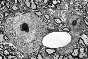
A nerve cell surrounded by axons and next to a capillary in a TEM section X2500
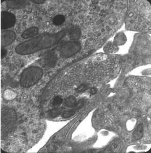
A synapse where one nerve cell contacts another type of nerve cell X30,000. Notice the tiny little round packets that contain the neural chemical messenger
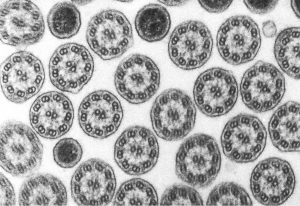
A field of cilia from a ciliated protozoan X50,000. Beating cilia propel small organisms along and help in feeding. Cilia are found in all complex cells, including many in human beings.