
Stereo imaging is where two images of the same subject are taken at a slightly different angle and then viewed separately in your two eyes. The brain is then forced to superimpose them to give a stereo version of the merged images. It was a popular thing to do in Victorian times to give more dimensionality to the pictures you are viewing. When applied to microscopy it gives an extra dimension to the images, but it can also provide depth information and be used for quantitative analysis of the relative depth of objects in a structure.
There are a number of ways to arrange your eyes to view the images. Stereo anaglyphs are where the two images are coloured red and green (sometimes blue or cyan). You then view them through red-green glasses, which are cheap and easy to purchase online. The red filter causes that eye to see the image dominated by the reddened colour, whilst the green filter causes the other eye to be dominated by the green image. The result is a 3D impression. An example shown here is a geranium leaf section taken in a scanning electron microscope.
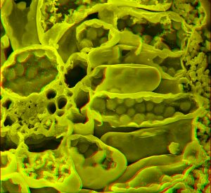
Leaf cells in stereo. Palisade cell on the right, mesophyll cell on the left and some of the leaf veins can be seen. The thick cell walls surround the cell, and round chloroplasts are visible inside (X1000).
Another way to merge the stereo images into one is with 3D glasses which focus one image into one eye and the other into the other eye. Some people can diverge their two eyes to create the same effect. The images below of nerve cells in a thick section of brain tissue have been obtained by using a microscope to create a stack of images at increasing depth through the section. The stack is then superimposed to create a 3D representation using software called PICOLAY (R) which allows the subject to be tilted. Either an anaglyph (upper image) or a stereopair (lower images) can then be created.
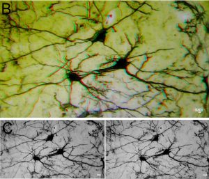
This network of brain cells has been stained using the method of Golgi, then a z-series (stack of images focussing down) has been created. By manipulating the stack, the subject can be tilted and reproduced either as an anaglyph (X500) or as a stereopair.
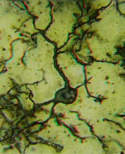
Anaglyph of an individual nerve cell with its spiny dendrites and round cell body, containing a central nucleus.
The same technique can be applied to buildings and other objects of any size. Below is a hydra, a small freshwater pond organism.
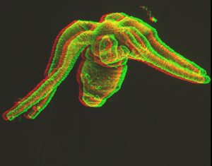
Hydra, a small (1 – 2 mm) tentacled organism from a pond *X50).
Stereopairs can also be taken in the transmission electron microscope. Here is an example showing spherical chromatin bodies inside the nucleus of a protozoan.
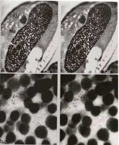
The upper pair shows a low power view of the nucleus of a protozoan (approx x2000), the lower pair a close up of the chromatin bodies in the nucleus. (approx X20,000).
And one last example……
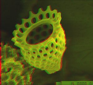
Anaglyph of a fossil radiolarian (X1000).