
The equipment used to examine samples consists of several optical microscopes, a transmission electron microscope (TEM), scanning electron microscope (SEM), and a confocal microscope. In addition, we have preparatory equipment (ultramicrotomes, gold coating unit etc) which we use to prepare the samples for microscopy.
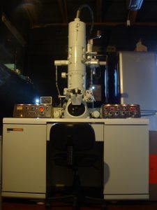
The TEM, a JEOL 100S model.
TEMs are used to image ultrathin sections, prepared by embedding fixed samples in a hard resin and then cutting them with an ultramicrotome to a typical thickness of 70 nm (see methods section).
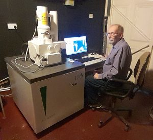
The SEM, a LEO 1430 model
SEM preparation typically involves drying a fixed sample through various methods and coating them with gold for placement in the microscope.
The confocal, a BioRad Radiance model
Confocal microscopy involves examining fluorescent samples (these can be naturally fluorescent) or samples stained using a technique that fluorescently labels individual proteins in a tissue or cell.