
Confocal microscopy is a technique for imaging fluorescence in a sample. Its power for biological applications lies in the ability to image fluorescently stained individual proteins in a tissue or cells using immunofluorescence methods. It can also be used for visualise other fluorescent molecules in various kinds of preparations.
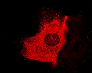
Immunostaining for the protein tubulin in a human cheek cell.
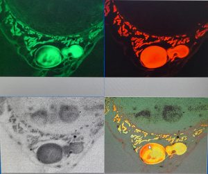
Fluorescent imaging of blood in an H&E stained earthworm section.
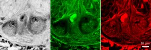
Fluorescent imaging of H&E stained earthworm nerve cord neurones (from the above section, seen at higher power.) Two neurones are seen in the bright field image on the left, with comparable fluorescent images in green and red channels on the right.