
Vorticella is a microscopic protozoan that has a bell-shaped body on a stalk that can contract. They can be found in many aquatic environments, and can be a pest in peoples’ aquaria. However, they are very attractive to look at, as you can see from the videos and images here.
Early microscopists called Vorticella the bell animalcule because of its shape. The stalk contracts into a spiral when the animal is disturbed as can be seen in some of the images below. The cilia form a ring around the mouth of the organism and when they beat, they produce a vortex in the fluid they live in, drawing food particles and prey items into their mouths (the name vorticella literally means ‘little vortex’).
A video of Vorticella in pond water.
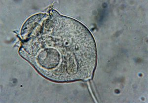
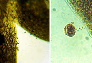
Light microscopic image of a living Vorticella. The cilia can be seen as hair-like structures surrounding the mouth. The circular body inside the animal is its contractile vacuole which drives out excess water.
A video of a living Vorticella (we recommend that you view it full screen; the coiling of the stalk can be seen in the lower right corner after it has contracted).
Because Vorticella is often attached to pond weed, as can be seen below, it is easy to harvest it and prepare it for electron microscopy.
A piece of pondweed with many vorticellids attached to it (X10)
For scanning electron microscopy, the weed is preserved in a fixative, then dehydrated gently and finally dried. There are a couple of different ways to dry it, depending on the quality you want to achieve. The sample below was the same piece of pondweed freeze-dried.
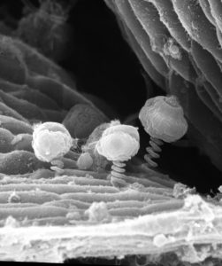
SEM of contracted Vorticella on pond weed. The stalk has contracted to a spring like appearance. X300 (approx).
With Vorticella living on a bit of pond weed, it can also be prepared for TEM quite easily. This involves embedding the weed with the Vorticella on it in a hard resin, then sectioning it very finely to look at in a TEM.
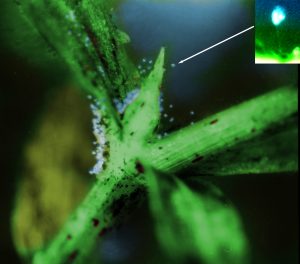
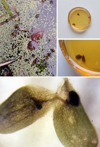
These images show duckweed in a pond which was found to have Vorticella on it. Pieces of the duckweed were fixed (see EM methods) and embedded in resin (two pictures on the right seen through a zoom microscope). The little dots in the lower panel are the Vorticella attached to the leaves of the duckweed. The images below that shows a close up of the colony of Vorticella and a single individual with its spiral stalk, both as seen within the resin.
Once embedded in resin, the protozoan can be sectioned thinly for the TEM. The image below is a cross section of the body of a Vorticella seen in ourTEM.
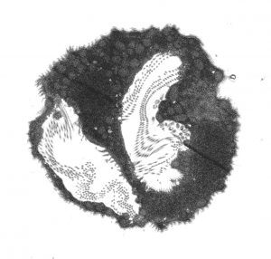
Low power TEM of the body of a Vorticella. The spiral-shaped mouth is sectioned across in two places (white areas) and is filled with the tiny cilia. X1000,
At very high magnification in a TEM, the structures of the cell and its components can be seen in detail down to the molecular level. Below are the tiny cilia and their basal bodies (roots) in a row.
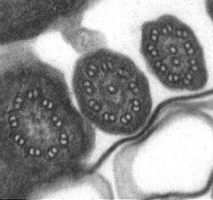
TEM of a cross section of a row of cilia , transitioning from the externally projecting shaft (on the right) into a basal body inside the body of the cell (on the left) as the section passes through different levels of the row . The ring-like structures are called microtubules. X250,000.