
Scanning electron microscopy is typically used to view the exterior of tissues and cells and it gives a great depth of focus, 3D view of your sample. However, it can also be used with a special adaptation of the methods, to view internal structures of cells – i.e. their organelles. Whilst cell internal structure was first visualised by transmission electron microscopy, methodologies were developed in the 1980s to provide internal views of cells (Tanaka K., Mitsushima A. (1984). A preparation method for observing intracellular structures by scanning electron microscopy. J. Microsc. 133, 213–222. 10.1111/j.1365-2818.1984.tb00487.x). This is not a commonly used technique, yet it offers incredible views of internal cell structure, as you can see with the examples below.
The basic structure of a cell is shown in a diagram based on TEM images. Below this are images showing the structures as seen in an SEM using a modification of Tanaka and Mitsushima’s method.
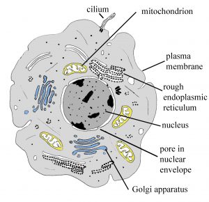
Diagram showing the ultrastructure of an animal cell based on TEM. This can be used to provide context for the EM images below.
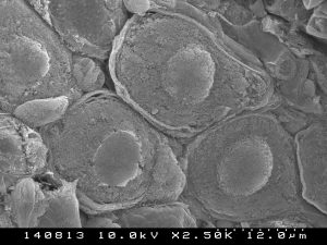
A group of nerve cells with central round nuclei, surrounded by cytoplasm, viewed by SEM. Uncoloured, original image, X2500.
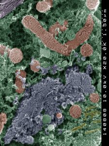
Cell organelles in the cytoplasm of a nerve cell. Mitochondria (red), Golgi body (blue) and endoplasmic reticulum (yellow) can be seen in the cytoplasm (green) in fabulous 3D detail. Colour enhanced SEM X20,000.
A mitochondrion from muscle.
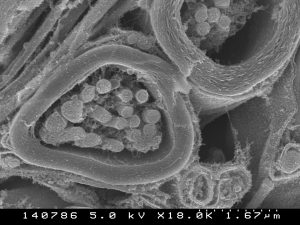
Cross section of a nerve fibre from the inner ear (X18,000).
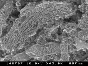
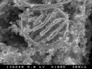
Golgi apparatus from a nerve cell (X45,000) .
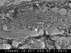
Endoplasmic reticulum and other organelles from a nerve cell (X20,000).